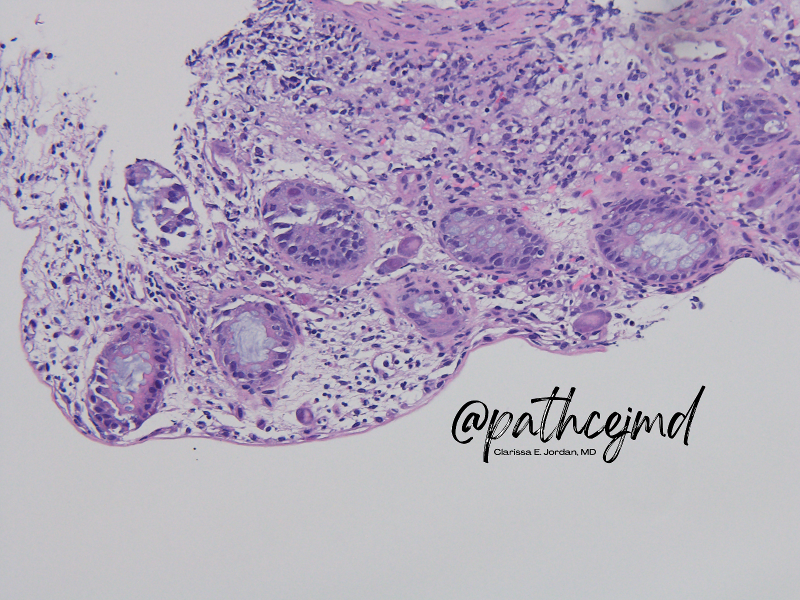
Case 7: CMV Colitis
Key features:
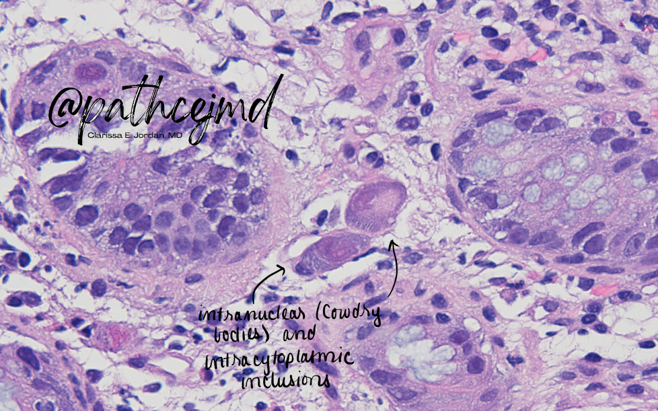
- CMV-infected cells are 2-4x larger than normal
- May contain Cowdry bodies (basophilic intranulcear inclusions surrounded by a clear halo)
- May contain smaller intracytoplasmic inclusions
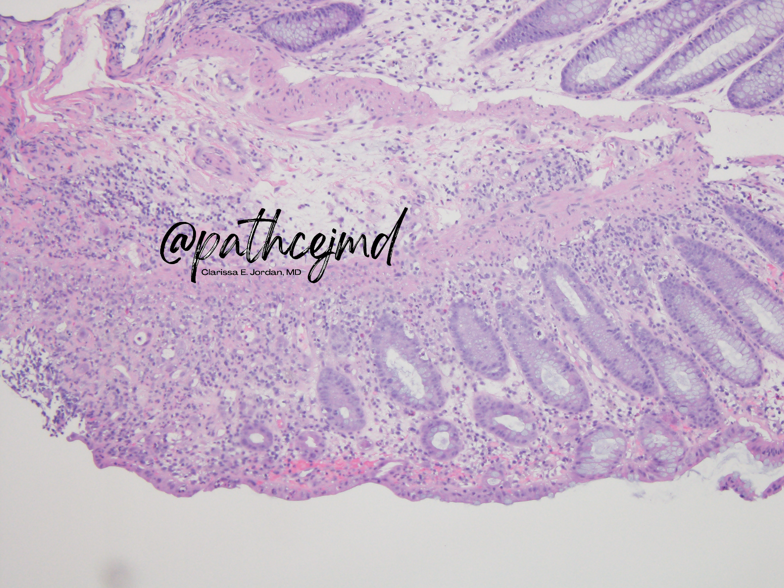
Features in background colon (non-specific):
- Architectural distortion
- Cryptitis
- Lymphoplasmacytic cellular infiltrates
- Reactive atypia
🦠 CMV has high prevalence in the general population (50-80% have abs by age 35)
Populations at risk for CMV disease (generally, immunocompromised folks), or those with:
- HIV (CD4 <50)
- Transplant recipients
- Heme malignancies
- Corticosteroid therapy
- Severe IBD
🦠 CMV initially infects mucosal epithelial cells, but stromal and vascular endothelial cells are the main targets in the GI tract
Inclusion bodies are rarely seen in epithelial cells around ulcer margins ➡️ common teaching of targeting ulcer base during endoscopic bx of suspected CMV dz
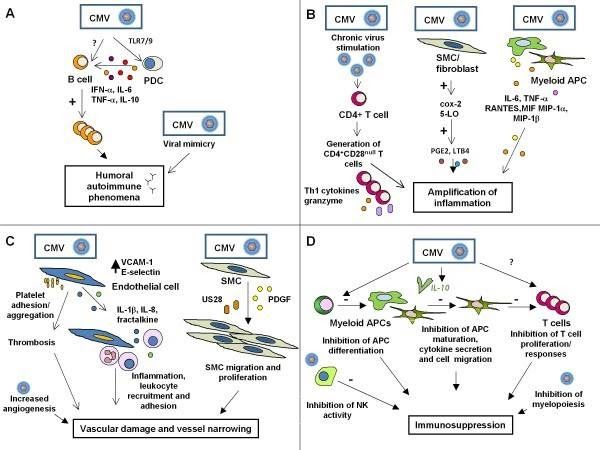
🦠 CMV has a huge array of effects on the immune system, and can lead to immunopaththologies, such as the amplification of inflammation and humoral autoimmune phenomenon,
See an excellent discussion of this ⬇️ by Varani and Landini https://pubmed.ncbi.nlm.nih.gov/21473750/
That’s all for this one! Thanks for following along 🤗
Special thanks to Dr. Roger Moreira, who I had the incredible privilege of learning from on this case
Find this case on Twitter:
#GIPath #Pathology case #tweetorial 🧵
— Clarissa E. Jordan, MD (@pathcejmd) March 9, 2022
Biopsies from a colonoscopy on a solid organ transplant recipient. What’s your diagnosis? (Poll ⬇️)#PathTwitter #MedEd @MayoClinicPath
(P.S. Always learning - I welcome any feedback!) pic.twitter.com/Fow6BtiTQh